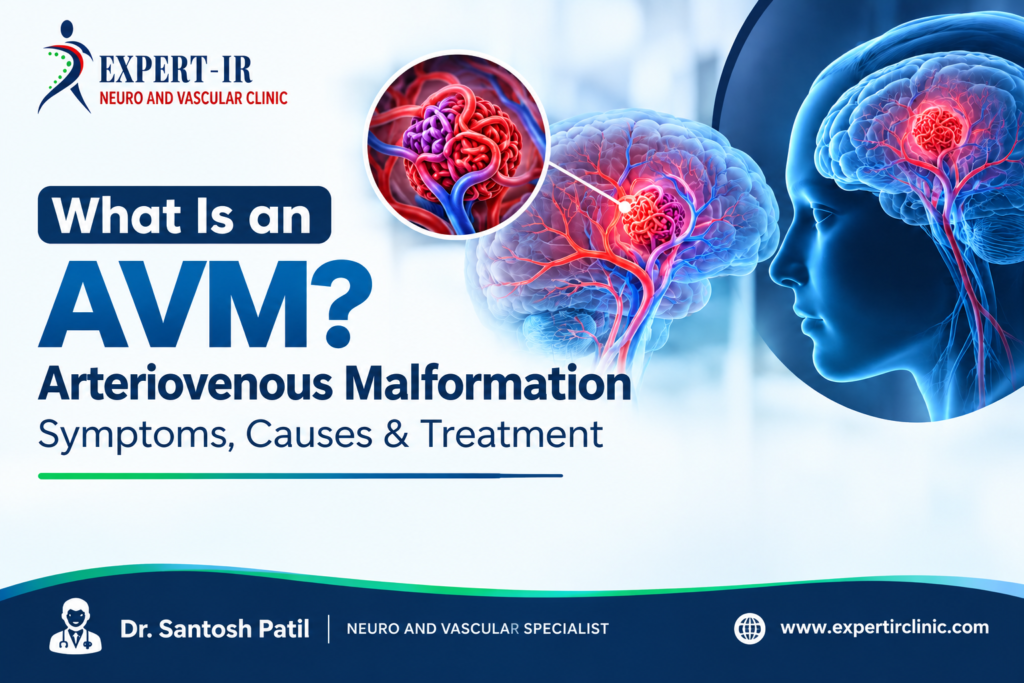
What Is an AVM? Arteriovenous Malformation Symptoms, Causes & Treatment
If you or someone you love has just heard the term AVM from a doctor, it’s natural to feel a little overwhelmed. Most people have never come across this condition before their own diagnosis, and the medical language around it can feel confusing at first. In simple terms, an arteriovenous malformation, or AVM, is an abnormal tangle of blood vessels that disrupts the normal flow of blood between arteries and veins. It can form almost anywhere in the body, though it is most often discussed in relation to the brain and spinal cord.
This article breaks down arteriovenous malformation symptoms, what causes an AVM, how doctors diagnose it, and the treatment options available today, including minimally invasive approaches like embolization. The goal is simple: help you understand what’s happening in the body, in plain language, so you can have a more informed conversation with your specialist.
What Is an AVM?
An arteriovenous malformation is a congenital condition, meaning it is typically present from birth, even though it may not cause any noticeable problems until later in life. Some people live for decades without ever knowing they have one.
Normally, blood flows from the heart through arteries, then through a network of tiny vessels called capillaries, and finally into veins to return to the heart. Capillaries slow the blood down and reduce pressure before it reaches the veins.
In an AVM, this capillary network is missing or poorly formed. Instead, arteries connect directly to veins. This creates a tangled cluster of vessels where high-pressure arterial blood flows straight into veins that are not built to handle that pressure.
How Does an AVM Affect Blood Flow?
Because the capillary buffer is missing, blood moves through an AVM faster and with more force than it should. Over time, this can lead to a few problems:
- The vein walls stretch and weaken because they are not designed for high-pressure blood flow.
- Surrounding tissue may not get enough oxygen, since blood bypasses the normal capillary exchange system.
- The fragile, abnormal vessels become more prone to leaking or rupturing.
This is essentially why AVMs are taken seriously by doctors, even when they are not currently causing symptoms. The structural weakness can build silently for years.
Common AVM Symptoms
AVM symptoms vary a lot depending on the size and location of the malformation. Some people never experience symptoms at all, and their AVM is only discovered during imaging done for an unrelated reason.
When symptoms do appear, they generally fall into two categories: general symptoms and location specific symptoms.
General Arteriovenous Malformation Symptoms
- Headaches that don’t respond well to usual pain relief
- Seizures, which are one of the most common early signs of a brain AVM
- Muscle weakness or numbness on one side of the body
- Vision changes or difficulty speaking
- Swelling, discoloration, or a warm patch of skin (more common with AVMs near the skin surface)
- A noticeable pulsing sound or feeling near the affected area
- Pain or a heavy, aching sensation in the affected limb or region
Signs of a Ruptured AVM
If an AVM bleeds or ruptures, symptoms tend to appear suddenly and should be treated as a medical emergency. These include a severe, sudden headache, confusion, loss of coordination, vision loss, or loss of consciousness.
Real world example: A patient in their late twenties with no prior health issues experienced a sudden, intense headache during a workout, followed by blurred vision. An emergency scan revealed a ruptured brain AVM. This is a fairly typical pattern seen in clinical practice, where the first noticeable symptom is also the most serious one.
Causes and Risk Factors
One of the most common questions patients ask is what caused my AVM. The honest answer is that researchers don’t fully understand why AVMs form.
Here’s what is currently known:
- AVMs are generally believed to develop during fetal development, meaning they are present at birth.
- They are not usually inherited, though rare genetic conditions can increase the likelihood of developing one.
- AVMs are not caused by lifestyle factors such as diet, exercise, or stress.
- They affect men and women at similar rates.
- Most AVMs are diagnosed between ages 20 and 40, though they can be found at any age.
Because there’s no clear preventable cause, doctors focus on early detection and monitoring rather than prevention strategies.
Is an AVM Dangerous?
This is understandably one of the biggest concerns for anyone recently diagnosed. The honest answer is that it depends on the size, location, and characteristics of the AVM.
Some AVMs remain stable for a person’s entire life and never cause a problem. Others carry a higher risk of bleeding, particularly if they are:
Located deep within the brain
Draining into a single vein rather than several
Associated with a nearby aneurysm
Discovered because of a previous bleed
A specialist will typically assess these risk factors before recommending observation, treatment, or ongoing monitoring. This is why an individualized evaluation matters so much. AVM risk is not the same from one patient to the next.
How Is AVM Diagnosed?
AVMs are usually found through imaging tests, either because a patient reports symptoms or because the AVM is discovered incidentally during a scan for something else.
Common diagnostic tools include:
MRI (Magnetic Resonance Imaging): Provides detailed images of soft tissue and blood vessels.
CT scan: Useful in emergencies to quickly check for bleeding.
Cerebral or spinal angiography: Considered the gold standard for mapping the exact structure of an AVM, since it shows blood flow in real time.
Doppler ultrasound: Sometimes used for AVMs closer to the skin’s surface, such as in limbs.
Once imaging confirms an AVM, doctors typically grade it using a scoring system that looks at size, location, and drainage pattern. This grading helps guide the treatment plan.
AVM Treatment Options
There is no single universal best AVM treatment. The right approach depends on the AVM’s size, location, symptoms, and overall risk profile. Specialists generally consider a few main paths:
1. Observation
For small, low-risk AVMs that aren’t causing symptoms, doctors may recommend regular monitoring through periodic imaging rather than immediate intervention.
2. Surgical Resection
In this approach, a surgeon physically removes the AVM. This tends to be most effective for AVMs located in accessible areas of the brain or body.
3. Stereotactic Radiosurgery
This is a non-invasive option that uses focused radiation beams to gradually shrink the AVM over months or even years. It is often used for smaller AVMs in locations that are hard to reach surgically.
4. Embolization
This minimally invasive procedure is increasingly used either as a standalone treatment or in combination with surgery or radiosurgery.
What Is AVM Embolization?
What Is AVM Embolization?
AVM embolization is a minimally invasive, image-guided procedure performed by an interventional specialist. Instead of open surgery, a thin catheter is guided through the blood vessels, usually starting from the groin or wrist, up to the site of the AVM.
Here’s a simplified step-by-step look at how it typically works:
Access: A small catheter is inserted into a blood vessel, usually in the leg or arm.
Navigation: Using real-time imaging, the doctor carefully guides the catheter through the vascular system to the exact location of the AVM.
Embolic agent delivery: A specialized material, such as a medical glue, coil, or particle, is released into the abnormal vessels to block off blood flow.
Closure: Once the AVM’s blood supply is reduced or cut off, the catheter is removed, and the small entry point is closed.
Embolization can shrink an AVM, reduce the risk of bleeding, or make it smaller and safer for a follow-up surgery or radiosurgery session. Recovery time is generally shorter than traditional open surgery, and many patients go home within a day or two, depending on the case.
Embolization isn’t always a standalone cure. In many cases, it works best as part of a combined treatment plan tailored to the specific AVM.
When Should You See a Specialist?
You should consult a vascular or interventional specialist if you experience:
Persistent or unusual headaches
Unexplained seizures
Sudden weakness, numbness, or vision changes
Visible swelling, discoloration, or a pulsing sensation under the skin
A family history of vascular malformations combined with any of the above symptoms
Early evaluation matters. Many AVM-related complications are manageable when caught and monitored before a rupture occurs.
Conclusion
An arteriovenous malformation is an abnormal connection between arteries and veins that can appear anywhere in the body, most notably in the brain and spine. While many AVMs stay silent for years, understanding the symptoms, risk factors, and available treatment paths, including minimally invasive options like embolization, can help patients and families make informed decisions with their care team.
Read More: Dr. Santosh Patil – Interventional Radiologist in Pune
Frequently Asked Questions (FAQs)
Prolonged sitting does not directly cause every case of varicose veins, but it reduces calf-muscle activity and can contribute to venous pooling and worsening symptoms, particularly when venous valve dysfunction already exists.
Prolonged sitting can reduce the calf-muscle pump and contribute to blood pooling in the legs. Recurrent evening swelling can also be a symptom of chronic venous insufficiency.
Avoid remaining completely stationary for prolonged periods. Getting up regularly to walk and performing ankle or calf movements during the workday can help maintain leg circulation.
Walking activates the calf-muscle pump and supports venous return. It can help reduce symptoms and support vein health, but it cannot repair already damaged vein valves.
A standing desk is not a complete solution. Prolonged standing can also increase venous pressure. The goal should be regular movement and changing positions, rather than replacing prolonged sitting with prolonged standing.
Some office workers may benefit from medical compression stockings, particularly if they experience leg swelling or heaviness. The appropriate compression level should be selected based on individual needs.
Exercise can improve circulation and reduce some symptoms, but established varicose veins caused by venous reflux generally do not disappear through exercise alone.
A venous Doppler ultrasound is commonly used to evaluate blood flow and identify venous reflux. It helps determine whether an underlying valve problem is responsible for the symptoms.
Consider an evaluation if you have persistent swelling, leg heaviness, aching, recurrent night cramps, increasingly prominent veins, skin changes, bleeding, or a wound around the ankle that is not healing.
Prolonged inactivity can aggravate symptoms and contribute to venous pooling. If significant venous reflux is already present, however, changing your sitting routine alone may not stop the underlying disease.
Age alone does not determine treatment suitability. If significant symptomatic venous reflux is confirmed, laser ablation may be considered after appropriate vascular assessment.